IV Contrast storage temperature


There are 2 different types of storage. One is storage of contrast in what I would call the supply room or storage room. For that, the temperature per Omnipaque product insert is 37°C . For current use contrast is placed in a contrast warmer where the temperature is 37 degrees celcius.
The temperature should no exceed 37°C after the closure has been entered.

For longer term storage the temperature is 27°C.

Benefits of Radiation


Many tests performed in the Department of Radiology and Biomedical Imaging use small amounts of ionizing radiation. Your doctor has requested these tests to better diagnose and manage your condition These tests are very different from radiotherapy in which much higher doses of ionizing radiation are used to treat cancers, because radiation can be targeted to kill cancers. Radiotherapy is performed by the Department of Radiation Oncology. One of the main benefits of radiation is that modern imaging allows for earlier and more accurate diagnosis, so that patients can be better treated and have better outcomes. For example, in a randomized study at Addenbrooke's Hospital in Cambridge, England, patients admitted because of severe abdominal pain were randomized to have a CT scan within 24 hours of admission or to have standard care. None of the 55 patients who got an early CT scan died, compared to 7 who died in the group of 63 patients who did not get an early CT scan . Such studies indicate the benefits of radiation through diagnostic tests are real and not just theoretical.

Software Tools to Limit Radiation Dose in CT


New software technology has also been introduced to help manage the noise from CT images, allowing for lower radiation dose. For instance, adaptive statistical iterative reconstruction (ASIR) may allow for dose reductions of up to 66% in abdominal scans, with no changes required to spatial or temporal resolution.21 One recent study compared CT enterography with ASIR with standard reconstruction, concluding that in patients weighing less than 160 lbs, examinations at 80 kVp with 30% ASIR produced diagnostically acceptable images. The average CTDIvol in the study was 6.15 mGy, and the average effective dose was 4.60 mSv.22 More generally, technologists can individualize the examination and adjust the radiation dose to the body type and body organ under examination, as the imaging of different body types and organs require different amounts of radiation. Prior to every CT examination, technologists should evaluate the appropriateness of the examination, and determine whether another type of examination may be more suitable. Higher resolution imaging is not always suitable for every given scenario. For instance, the likelihood of detection of small pulmonary masses may not be increased with higher resolution imaging.
Ultimately, technologists and radiologists, as well as the referring clinicians ordering CT images, required an increased level of education to understand the risks and limitations of CT, as well as the clinical applications that are likely to provide the greatest benefit.

The CT Technologist's Role in Limiting Radiation Dose


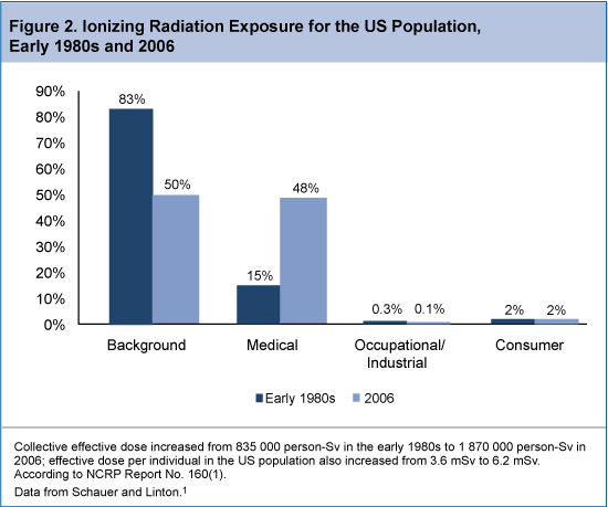
 The CT technologist should be aware of the radiation risks associated with CT, and can play a leadership role in implementing tools to limit radiation exposure in patients undergoing CT imaging. Specifically, CT technologists need to understand the parameters that affect radiation dose, practice dose reduction techniques on a regular basis, and be aware of the CT examination types that are the biggest contributors to radiation exposure from CT imaging. The increased use of CT in routine clinical practice has likely resulted in a substantial increase in radiation exposure in the US population. In fact, a report from the National Council on Radiation Protection and Measurements noted as much as 7-fold increase in ionizing radiation exposure from medical procedures between the early 1980s and 2006 (Figure 2). The report estimated that in 2006, 48% of total radiation exposure was from medical procedures, as opposed to just 15% in the early 1980s.13 Among the leading contributors to the increase in CT-associated radiation exposure include imaging studies of the head, chest, abdomen/pelvis, and studies of the chest for the diagnosis of pulmonary embolism.
The ongoing desire to limit radiation exposure must be balanced with the need to obtain adequate images during CT examinations. In general, higher radiation doses result in higher-resolution images, whereas lower doses lead to increased image noise and less sharp images. However, increased radiation dosage increases the risk of adverse side effects, most notably the risk of radiation-induced cancer. For instance, a 4-phase abdominal CT gives the same radiation dose as 400 chest X-rays. Fortunately, multiple strategies exist that can reduce the exposure to ionizing radiation during a CT scan.
Computed tomography technologists can employ a variety of dose reduction techniques that adequately balance the need for adequate imaging with adequate patient protection. For instance, technologists can control the z-axis scan length or minimize the number of phases to limit radiation exposure. External bismuth body shields to the breast, gonads, and thyroid are commonly used to protect these sensitive areas during CT imaging. In addition, dose reduction hardware techniques are commonly employed, including noise index and automatic tube current modulation, adjustments for peak kilovoltage (kVp), rotation time, and precise centering on the area to be scanned. In one analysis of the use of bismuth shielding, organ-based tube current modulation, and global tube current reduction as radiation dose reduction strategies using thorax phantoms of different sizes (15, 30, 35, and 40 cm lateral width), radiation dose to the breast region was reduced by approximately 21% and 37% with pediatric (2-ply shield; 15-cm phantom) and adult (4-ply shield; 30-, 35-, and 40-cm phantoms) models, respectively. Organ-based tube current modulation achieved dose decreases of 12% in the pediatric model and 34% to 39% in the adult model, and global lowering of the tube current reduced breast dosage by 23% and 39% in the pediatric and adult models, respectively.

The Importance of Patient Centering to Limit Radiation Dose in CT


The use of various tools that influence radiation dose and image noise, such as X-ray intensity shaping filters (bowtie filters), often requires accurate patient centering in the scan field of view. With the use of these bowtie filters, patient centering is critical to achieve the optimal balance of reduced radiation dose with reduced noise. To assess the influence of patient centering on image quality and dose requirements, researchers conducted imaging scans with bowtie filters using phantom centers that were positioned at 0, 3, and 6 cm below the center of rotation, or isocenter. These models were then retrospectively applied to scans from 273 adult body patients. The researchers reported that miscentering by 3 to 6 cm resulted in an increase in surface dose of 18% and 41%, respectively, and an increase in image noise of 6% and 22%, respectively. In addition, the retrospective analysis of adult body scout scan projection radiograph scans found that 46% of patients were miscentered in elevation by 20 to 60 mm, with a mean of 23 mm below the isocenter. Overall, the study concluded that patient miscentering resulted in a dose penalty of up to 140%, with a mean penalty of 33%, assuming that increases in tube current were necessary to compensate for increased image noise. The authors suggested automatically providing patient-specific centering and scan parameter selection information during CT studies so that radiologic technologists could improve workflow, achieve consistent image quality, and reduce radiation dose.19
Another study focused on the impact of patient miscentering on image noise and surface radiation dose using 3 different commercial CT scanners. Phantoms were positioned at 0, 2, 4, and 6 cm below the isocenter of the scanner's field of view. The resulting measurements were applied to imaging studies from 480 patients. The researchers reported that for a 64-slice CT scanner, the maximum increase in surface dose was 13.5%, 33.3%, and 51.1% with miscenterings of 2, 4, and 6 cm, respectively. An analysis of the available patient scout scans determined that patients were miscentered by an average of 2.2 cm below the isocenter. This resulted in increases in patient dose and image noise by 23% and 7%, respectively.20These studies demonstrate the importance of patient centering to limit radiation dose while optimizing image quality.

CT Parameters Affecting Radiation Dose


The discussion of strategies to estimate and limit radiation dose in patients undergoing CT will require a review of the specific CT parameters that most affect radiation dose, as defined in the sections below.8
Kilovolts
Kilovolt (kV) is the amount of voltage between an X-ray tube's anode and cathode. It determines the energy of the X-ray being emitted. Higher energy X-rays have a greater potential of passing through the body and creating a signal at the detector than lower energy X-rays. Higher kV also means less noise. High-energy X-rays are absorbed by the body and deposit more energy than low-energy X-rays; therefore, contribute more to patient dose. For example, changing the kV from 120 to 135 increases the radiation dose delivered by approximately 33%. However, higher kV is sometimes warranted in larger patients or dense anatomy such as bone where increased penetration is needed.
MilliAmpere SecondsThe tube current or milliAmpere seconds (mAs) determines the number of X-rays the tube produces. Combined with the gantry rotation time, mAs represents the total X-ray output of the tube per rotation. Changing the mAs, is the most common method of adjusting dose and noise level. Cutting the mAs in half will reduce the dose by a factor of 2; however, it will also half the number of X-rays reaching the detector. Because fewer X-rays are detected, the image noise will increase and low contrast detectability will be diminished.
CT Pitch and Helical Pitch-Beam Pitch
CT pitch and helical pitch-beam pitch is defined as the distance the table travels in a rotation divided by the total active detector width in the Z direction. Helical pitch is the same except it is divided by the individual channel thickness rather than the total collimation. The higher the CT helical pitch, the faster the table moves through the X-ray beam. This results in lower radiation dose to the patient, however it may result in increased noise and may cause helical artifact. One may need to increase mAs as the pitch increases to maintain image quality.
Effective mAs Effective mAs (mAs eff) is the mAs divided by the pitch. Because pitch affects the patient dose, mAs by itself does not completely represent the number of X-rays entering the patient.
CollimationWith multislice scanning, there are many combinations of slice width and the number of slices that may be used to acquire the scan volume. With all collimators on multislice systems, the actual X-ray beam is slightly wider than the nominal beam width. This is to ensure that the detectors on the edge of the array receive uniform X-ray coverage. This results in a small amount of unused radiation. For the best image quality and dose efficiency, using the thinnest slices to cover the entire detector is ideal.
Acquired and Reconstructed Slice WidthAcquired is the actual slice thickness at which the scan was taken. Reconstructed slice width refers to post processing of the acquired slice. The acquired slice width is governed by the multislice detector configuration and determines the minimum image width that can be reconstructed. Thicker reconstructed images have less noise with all other factors being equal. However, thicker slices have less resolution. Scanning with the thinnest possible slice avoids partial volume artifacts and allows flexibility for excellent multiplanar reformats and 3-dimensional rendering without higher radiation dose. The optimal image quality and dose efficiency are achieved with the thinnest slice covering the entire detector.
Reconstruction KernelThe acquired data are filtered by the reconstruction kernel. The kernel plays a large role in determining spatial resolution. This has a great effect on the amount of noise in the image and the dose needed for a given level of image noise. There are a large variety of reconstruction kernels to select. The choice of the kernel is made based on the clinical need. Sharper reconstruction kernels are used for better in-plane spatial resolution such as in the lungs to help identify tiny airways. Smooth reconstruction kernels reduce the image noise at the expense of in-plane resolution; they are used in body imaging to reduce noise and enhance contrast resolution.
Detector Efficiency
Detector efficiency has a huge influence on the radiation dose delivered during a CT examination. The detector's ability to capture the X-ray and convert it to light, then transmit that light and convert it into an electrical signal with minimal loss, defines the overall efficiency of the detector. Better efficient detectors result in lower patient doses for a given level of image quality.
FiltrationFiltration is added outside the X-ray tube to block low-energy X-rays (photons) and reduce patient dose. The lowest energy photons will not pass through the body at all and will only contribute to patient dose. In the process of removing these low-energy photons, some desirable medium- and high-energy X-rays will be removed as well. This results in decreasing overall output of the X-ray tube. An increase in tube current or mAs may be needed to maintain image quality. A CT system needs enough filtration to block the lowest energy photons but not so much to lose the ability to distinguish low-contrast anatomy.
Patient SizeThe size of the patient plays a significant role in the total dose absorbed for the same technique. It is important to tailor the kV and mAs to the patient size to minimize the dose to the patient. With smaller patients, such as pediatric patients, the dose can be 2 to 3 times higher than on an adult. Lower kV and mAs should be used to achieve the small image quality.
Body Part Being ScannedDifferent organs in the body have different sensitivities to radiation (Table 1). For instance, the lungs are not as dense as the abdomen or pelvis because they are air filled. Therefore, less radiation (kVp and mA) is needed for imaging.
AgeThe risk of developing cancer from a CT examination decreases with increased age. Younger patients' organs are more radiosensitive in general, due to the rapid rate of cell division and growth at a young age. It is critical in young patients to keep the dose as low as possible while maintaining image quality.

How radiation affects cells


The primary way radiation affects our health is through breakage of DNA molecules. DNA is a long chain of amino acids whose pattern forms the blueprint on how the cell lives and functions.  Radiation is able to break that chain. When it does,  three things can happen:  

1) The DNA is repaired properly

In this case, the cell is repaired properly and it continues to function normally. DNA breakage occurs normally every second of the day and cells have a natural ability to repair that damage.

2) The DNA damage is so severe that the cell dies (deterministic effects)

When the DNA or other critical parts of a cell receive a large dose of radiation, the cell may either die or be damaged beyond repair. If this happens to a large number of cells in a tissue or organ, early radiation effects may occur. These are called deterministic effects and the severity of the effects varies according to the radiation dose received. They can include burns, cataracts, and in extreme cases, death.

The first evidence of deterministic effects became apparent with early experimenters and users of radiation. They suffered severe skin and hand damage due to excessive radiation dose. More recently, this relationship was observed at the 1986 Chernobyl nuclear plant accident where more than 130 workers and firefighters received high radiation doses (800 to 16,000 mSv), and suffered severe radiation sickness. Two of the people exposed died within days of exposure. Close to 30 more workers and firefighters died within the first three months.

The CNSC and other international regulators put measures in place, including stringent dose limits and radioactive source tracking databases, to mitigate the chances of the public or workers receiving doses of radiation high enough to cause deterministic effects. The CNSC also has strict regulations on how nuclear substances and devices must be handled in Canada. 
3) The cell incorrectly repairs itself, but it continues to live (stochastic effects)

In some cases, the DNA of the cell may be damaged by radiation, but the damage does not kill the cell.  The cell may continue to live and even reproduce itself, but the cell and its descendents may no longer function properly and may disrupt the function of other cells.  The probability of this type of  detrimental effect is proportionate to the dose and it is called a stochastic effect – when there is a statistical probability that the effects of exposure will occur. In such cases, the likelihood of the effects increases as the dose increases. However, the timing of the effects or their severity does not depend on the dose. 

This process happens all the time in everyone. In fact, people are exposed to about 15,000 such events every second of every day. Sometimes, the cell structure changes because it repairs itself improperly. This alteration could have no further effect, or the effect could show up later in life. Cancer and hereditary effects may or may not take place.

Radiation-induced hereditary effects


Genetic damage occurs when the DNA of sperm or egg cells are damaged. This causes a harmful characteristic that is passed on from one generation to the next. Animal studies, such as those conducted on fruit flies by Hermann J. Muller in 1926, showed that radiation will cause genetic mutations. However, to date there have been no known genetic effects in humans caused by radiation. This includes studies involving some 30,000 children of survivors of the atomic bombings of the cities of Hiroshima and Nagasaki in Japan in 1945 (BEIR VII).

Broadcast Antennas Radiation exposure


Radio and television broadcast stations transmit their signals via RF electromagnetic waves. Broadcast stations transmit at various RF frequencies, depending on the channel, ranging from about 550 kHz for AM radio up to about 800 MHz for some UHF television stations. Frequencies for FM radio and VHF television lie in between these two extremes. Operating powers can be as little as a few hundred watts for some radio stations or up to millions of watts for certain television stations. Some of these signals can be a significant source of RF energy in the local environment, and the FCC requires that broadcast stations submit evidence of compliance with FCC RF guidelines. 
The amount of RF energy to which the public or workers might be exposed as a result of broadcast antennas depends on several factors, including the type of station, design characteristics of the antenna being used, power transmitted to the antenna, height of the antenna and distance from the antenna. Since energy at some frequencies is absorbed by the human body more readily than energy at other frequencies, the frequency of the transmitted signal as well as its intensity is important.

Public access to broadcasting antennas is normally restricted so individuals cannot be exposed to high-level fields that might exist near antennas. Measurements made by the FCC, EPA, and others have shown that ambient RF radiation levels in inhabited areas near broadcasting facilities are typically well below the exposure levels recommended by current standards and guidelines. Antenna maintenance workers are occasionally required to climb antenna structures for such purposes as painting, repairs, or beacon replacement. Both the EPA and OSHA have reported that in these cases it is possible for a worker to be exposed to high levels of RF energy if work is performed on an active tower or in areas immediately surrounding a radiating antenna. Therefore, precautions must be taken to ensure that maintenance personnel are not exposed to unsafe RF fields.

Portable Radio Systems Radiation exposure


"Land-mobile" communications include a variety of communications systems that require the use of portable and mobile RF transmitting sources. These systems operate in narrow frequency bands between about 30 and 1,000 MHz. Radio systems used by the police and fire departments, radio paging services, and business radio are a few examples of these communications systems. There are essentially three types of RF transmitters associated with land-mobile systems: base-station transmitters, vehicle-mounted transmitters, and handheld transmitters. The antennas used for these various transmitters are adapted for their specific purpose. For example, a base-station antenna must radiate its signal to a relatively large area, and, therefore, its transmitter generally has to use higher power levels than a vehicle-mounted or handheld radio transmitter. Although these base-station antennas usually operate with higher power levels than other types of land-mobile antennas, they are normally inaccessible to the public since they must be mounted at significant heights above ground to provide for adequate signal coverage. Also, many of these antennas transmit only intermittently. For these reasons, such base-station antennas have generally not been of concern with regard to possible hazardous exposure of the public to RF radiation. Studies at rooftop locations have indicated that high-powered paging antennas may increase the potential for exposure to workers or others with access to such sites, for example, maintenance personnel. Transmitting power levels for vehicle-mounted land-mobile antennas are generally less than those used by base-station antennas but higher than those used for handheld units.

Handheld portable radios such as walkie-talkies are low-powered devices used to transmit and receive messages over relatively short distances. Because of the low power levels used, the intermittence of these transmissions, and the fact that these radios are held away from the head, they should not expose users to RF energy in excess of safe limits. Therefore, the FCC does not require routine documentation of compliance with safety limits for push-to-talk two-way radios.

Radar Systems Radiation exposure


Radar systems detect the presence, direction, or range of aircraft, ships, or other moving objects. This is achieved by sending pulses of high-frequency electromagnetic fields (EMF). Radar systems usually operate at radiofrequencies between 300 megahertz (MHz) and 15 gigahertz (GHz). Invented some 60 years ago, radar systems have been widely used for navigation, aviation, national defense, and weather forecasting. People who live or routinely work around radar have expressed concerns about long-term adverse effects of these systems on health, including cancer, reproductive malfunction, cataracts, and adverse effects for children. It is important to distinguish between perceived and real dangers that radar poses and to understand the rationale behind existing international standards and protective measures used today.

The power that radar systems emit varies from a few milliwatts (police traffic-control radar) to many kilowatts (large space tracking radars). However, a number of factors significantly reduce human exposure to RF generated by radar systems, often by a factor of at least 100:


  • Radar systems send electromagnetic waves in pulses and not continuously. This makes the average power emitted much lower than the peak pulse power.
  • Radars are directional and the RF energy they generate is contained in beams that are very narrow and resemble the beam of a spotlight. RF levels away from the main beam fall off rapidly. In most cases, these levels are thousands of times lower than in the main beam.
  • Many radars have antennas which are continuously rotating or varying their elevation by a nodding motion, thus constantly changing the direction of the beam.
  • Areas where dangerous human exposure may occur are normally inaccessible to unauthorized personnel.

Symptoms Radiation sickness


The severity of signs and symptoms of radiation sickness depends on how much radiation you've absorbed. How much you absorb depends on the strength of the radiated energy and the distance between you and the source of radiation. Signs and symptoms also are affected by the type of exposure — such as total or partial body and whether contamination is internal or external — and how sensitive to radiation the affected tissue is. For instance, the gastrointestinal system and bone marrow are highly sensitive to radiation.
Absorbed dose and duration of exposure
The absorbed dose of radiation is measured in a unit called a gray (Gy). Diagnostic tests that use radiation, such as an X-ray, result in a small dose of radiation — typically well below 0.1 Gy, focused on a few organs or small amount of tissue.
Signs and symptoms of radiation sickness usually appear when the entire body receives an absorbed dose of at least 1 Gy. Doses greater than 6 Gy to the whole body are generally not treatable and usually lead to death within two days to two weeks, depending on the dose and duration of the exposure.
Initial signs and symptoms
The initial signs and symptoms of treatable radiation sickness are usually nausea and vomiting. The amount of time between exposure and when these symptoms develop is an indicator of how much radiation a person has absorbed.
After the first round of signs and symptoms, a person with radiation sickness may have a brief period with no apparent illness, followed by the onset of new, more serious symptoms.
In general, the greater your radiation exposure, the more rapid and more severe your symptoms will be.
Early symptoms of radiation sickness*
 Mild exposure (1-2 Gy)Moderate exposure (2-6 Gy)Severe exposure (6-8 Gy)Very severe exposure (8-10 Gy or higher)
Nausea and vomitingWithin 6 hoursWithin 2 hoursWithin 1 hourWithin 10 minutes
Diarrhea--Within 8 hoursWithin 3 hoursWithin 1 hour
Headache--Within 24 hoursWithin 4 hoursWithin 2 hours
Fever--Within 3 hoursWithin 1 hourWithin 1 hour
Later symptoms of radiation sickness*
Dizziness and disorientation-- --Within 1 weekImmediate 
Weakness, fatigue Within 4 weeksWithin 1-4 weeksWithin 1 weekImmediate 
Hair loss, bloody vomit and stools, infections, poor wound healing, low blood pressure--Within 1-4 weeksWithin 1 weekImmediate
* Adapted from Radiation exposure and contamination. The Merck Manuals: The Merck Manual for Healthcare Professionals.
When to see a doctor
An accident or attack that causes radiation sickness would no doubt cause a lot of attention and public concern. If such an event occurs, monitor radio, television or online reports to learn about emergency instructions for your area.
If you know you've been exposed to radiation, seek emergency medical care.

Treatments and drugs of Radiation Sickness


The treatment goals for radiation sickness are to prevent further radioactive contamination; treat life-threatening injuries, such as from burns and trauma; reduce symptoms; and manage pain.
Decontamination
Decontamination is the removal of as much external radioactive particles as possible. Removing clothing and shoes eliminates about 90 percent of external contamination. Gently washing with water and soap removes additional radiation particles from the skin.
Decontamination prevents further distribution of radioactive materials and lowers the risk of internal contamination from inhalation, ingestion or open wounds.
Treatment for damaged bone marrow
A protein called granulocyte colony-stimulating factor, which promotes the growth of white blood cells, may counter the effect of radiation sickness on bone marrow. Treatment with this protein-based medication, which includes filgrastim (Neupogen) and pegfilgrastim (Neulasta), may increase white blood cell production and help prevent subsequent infections.
If you have severe damage to bone marrow, you may also receive transfusions of red blood cells or blood platelets.
Treatment for internal contamination
Some treatments may reduce damage to internal organs caused by radioactive particles. Medical personnel would use these treatments only if you've been exposed to a specific type of radiation. These treatments include the following:
  • Potassium iodide. This is a nonradioactive form of iodine. Because iodine is essential for proper thyroid function, the thyroid becomes a "destination" for iodine in the body. If you have internal contamination with radioactive iodine (radioiodine), your thyroid will absorb radioiodine just as it would other forms of iodine. Treatment with potassium iodide may fill "vacancies" in the thyroid and prevent absorption of radioiodine. The radioiodine is eventually cleared from the body in urine. Potassium iodide isn't a cure-all and is most effective if taken within a day of exposure.
  • Prussian blue. This type of dye binds to particles of radioactive elements known as cesium and thallium. The radioactive particles are then excreted in feces. This treatment speeds up the elimination of the radioactive particles and reduces the amount of radiation cells may absorb.
  • Diethylenetriamine pentaacetic acid (DTPA). This substance binds to metals. DTPA binds to particles of the radioactive elements plutonium, americium and curium. The radioactive particles pass out of the body in urine, thereby reducing the amount of radiation absorbed.
Supportive treatment
If you have radiation sickness, you may receive additional medications or interventions to treat:
  • Bacterial infections
  • Headache
  • Fever
  • Diarrhea
  • Nausea and vomiting
  • Dehydration
  • Burns
End-of-life care
A person who has absorbed large doses of radiation (6 Gy or greater) has little chance of recovery. Depending on the severity of illness, death can occur within two days or two weeks. People with a lethal radiation dose will receive medications to control pain, nausea, vomiting and diarrhea. They may also benefit from psychological or pastoral care.

Tests and diagnosis of Radiation Sickness


When a person has experienced known or probable exposure to a high dose of radiation from an accident or attack, medical personnel take a number of steps to determine the absorbed radiation dose. This information is essential for determining how severe the illness is likely to be, which treatments to use and whether a person is likely to survive.

Information important for determining an absorbed dose includes:

  • Known exposure. Details about distance from the source of radiation and duration of exposure can help provide a rough estimate of the severity of radiation sickness.
  • Vomiting and other symptoms. The time between radiation exposure and the onset of vomiting is a fairly accurate screening tool to estimate absorbed radiation dose. The shorter the time before the onset of this sign, the higher the dose. The severity and timing of other signs and symptoms may also help medical personnel determine the absorbed dose.
  • Blood tests. Frequent blood tests over several days enable medical personnel to look for drops in disease-fighting white blood cells and abnormal changes in the DNA of blood cells. These factors indicate the degree of bone marrow damage, which is determined by the level of an absorbed dose.
  • Dosimeter. A device called a dosimeter can measure the absorbed dose of radiation but only if it was exposed to the same radiation event as the affected person.
  • Survey meter. A device such as a Geiger counter can be used to survey people to determine the body location of radioactive particles.
  • Type of radiation. A part of the larger emergency response to a radioactive accident or attack would include identifying the type of radiation exposure. This information would guide some decisions for treating people with radiation sickness.

Application of the Basic Standards in Radiation Safety


The BSS set out detailed requirements for practices and interventions to protect workers, patients and the general public from radiation exposure. They also recommend procedures for ensuring the safety of sources, for accident prevention, for emergency planning and preparedness and for mitigating the consequences of accidents. Although the majority are of a qualitative nature, the BSS also establish many requirements expressed in terms of restrictions or guidance on the dose that may be incurred by people. The range of doses spreads over four orders of magnitude, from ones that are so minute that they should be exempt from the requirements to doses that are large enough to make intervention almost mandatory.
National governments usually have the responsibilities for enforcing radiation safety standards, generally through a system that includes a regulatory authority. In addition, governments usually provide for certain essential services for radiation protection and safety and for interventions that exceed or that complement the capabilities of regulators. The BSS can only be effectively applied when such a national infrastructure is firmly in place. In addition to legislation and regulations, the essential elements are:
A Regulatory Authority. This should be empowered to authorize and inspect, and to enforce the legislation and regulations. It must have sufficient resources, including adequate numbers of trained personnel. There must be arrangements for detecting any build up of radioactive substances in the general environment, for disposing of radioactive waste and preparing for interventions, particularly during emergencies, that could result in exposure of the public.
Education, training and public information. There must be adequate arrangements and resources for these, as well as for the exchange of information among specialists. There must also be appropriate means of informing the public, its representatives and the information media about health and safety concerns.
Facilities and services for radiation protection and safety must be well established at the national level. These include laboratories for personal dosimetry and environmental monitoring, and calibration and intercomparison of radiation measuring equipment; they could also include central registries for radiation dose records and information on equipment reliability.
To ensure radiation safety, the BSS promotes development of:
  • A Safety Culture - that encourages a questioning and learning attitude to protection and safety, and discourages complacency.
  • Quality Assurance Programmes - that provide, as appropriate, adequate assurances that the specified requirements related to protection and safety are satisfied.
  • Control of Human Factors - limiting, as far as practicable, the contribution of human error to accidents and other events that could give rise to exposures. This can be achieved by ensuring that all personnel on whom protection and safety depend are appropriately trained and qualified.
  • Qualified experts - made available for providing advice on the observance of the BSS.
The BSS promotes sound technical planning and implementation through the following:
Security of sources. Radiation sources must be kept secure so as to prevent theft or damage.
Defence in depth. A multilayer system of protection and safety provisions commensurate with the radiation hazards involved is applied to sources, so that a failure at one layer is compensated for or corrected by subsequent layers.
Good engineering practice. This reflects approved codes and standards, and must be supported by reliable management and organization to ensure protection and safety throughout the life of the sources.
Verification of safety. Protection and safety measures for sources must be made in a way that they can be regularly monitored and verified for compliance. In addition, records should be kept of the results of monitoring and verification.
Transport
Additionally, radioactive substances have to be transported in accordance with the IAEA Regulations for the Safe Transport of Radioactive Material and with any applicable international convention.
Under the BSS, interventions apply to the following:
  • Emergencies, where protective action is needed to reduce or avert temporary radiation exposures, including accidents at nuclear installations (for which emergency plans or procedures have been activated).
  • Chronic exposure situations requiring remedial action to reduce or avert long-term radiation exposure. This includes exposure to radon in buildings and exposure to radioactive residues from past events.
        
Exposure resulting fromBasisEquivalent period of global exposure to average natural background

Nuclear weapons testingAll past tests2- 3 years
Apparatus and substances used in medicineOne year of pracitce at the current rate90 days
Severe accidentsAccidents to date20 days
Nuclear power generation (under normal operating conditions)Total nuclear generation to date. One year of practice at the current rate1 day
Occupational activitiesOne year of occupational activities at the current rate8 hours

The table presents the UNSCEAR summary of the relative radiological impact from some practices as well as from severe accidents that required intervention. The levels of radiation exposure are expressed as equivalent periods of exposures to natural resources.

Sources of Artificial Radiation


Doses from artificial radiation are, for most of the population, much smaller than those from natural radiation but they still vary considerably. They are in principle fully controllable, unlike natural sources.
Medical. Radiation is used in medicine in two distinct ways: to diagnose disease or injury; and to kill cancerous cells. In the oldest and most common diagnostic use, X rays are passed through the patient to produce an image. The technique is so valuable that millions of X ray examinations are conducted every year. One chest X ray will give 0.1 mSv of radiation dose. For some diseases, diagnostic information can be obtained using gamma rays emitted by radioactive materials introduced into the patient by injection, or by swallowing or by inhalation. This technique is called nuclear medicine. The radioactive material is part of a pharmaceutical chosen so that it preferentially locates in the organ or part of the body being studied. To follow the distribution or flow of the radioactive material a gamma camera is used. It detects the gamma radiation and produces an image, and this indicates whether the tissue is healthy or provides information on the nature and extent of the disease.
Cancerous conditions may be treated through radiotherapy, in which beams of high energy X rays or gamma rays from cobalt-60 or similar sources are used. They are carefully aimed to kill the diseased tissue, often from several different directions to reduce the dose to surrounding healthy tissue. Radioactive substances, either as small amounts of solid material temporarily inserted into tissues or as radioactive solutions, can also be used in treating diseases, delivering high but localised radiation doses.
Medical uses of radiation are by far the largest source of man-made exposure of the public; the global yearly average dose is 0.3 millisieverts.
Environmental Radiation. Radioactive materials are also present in the atmosphere as a result of atomic bomb testing and other activities. They may lead to human exposure by several pathways external irradiation from radioactive materials deposited on the ground; inhalation of airborne radioactivity, and ingestion of radioactive materials in food and water.
Radioactive fall-out from nuclear weapons tests carried out in the atmosphere is the most widespread environmental contaminant but doses to the public have declined from the relatively high values of the early 1960s to very low levels now. The global yearly average dose is 0.006 millisieverts. However, where tests were carried out at ground level or even underground, localised contamination often remains near weapons sites.
Nuclear and other industries, and to a small degree hospitals and universities, discharge radioactive materials to the environment. Nearly all countries regulate industrial discharges and require the more significant to be authorized and monitored. Monitoring of such effluent may be carried out by the government department that authorizes the discharges as well as by the operator.
The nuclear power industry releases small quantities of a wide variety of radioactive materials at each stage in the nuclear fuel cycle. For the public the global yearly average dose is 0.008 millisieverts. The type of radioactive materials, and whether they are liquid, gaseous or particulate depends upon the operation of each process. For instance, nuclear power stations release carbon-14 and sulphur-35, which find their way through food chains to humans. Liquid discharges include radioactive materials that people may ingest through fish and shellfish.
The yearly dose to individuals living close to a power plant is small - usually a fraction of a millisievert; doses to people further away are even smaller. Reprocessing nuclear fuel produces higher doses which vary greatly from plant to plant. For the most exposed members of the public, they can be as high as 0.4 millisieverts, but for most of the population they are very much smaller.
World-wide, there are estimated to be four million workers exposed to artificial radiation as a result of their work, with an average yearly dose of about 1 millisievert. Another five million (mostly in civil aviation) have yearly average doses due to natural radiation of 1.7 millisieverts.
Non-nuclear industries also produce radioactive discharges. They include the processing of ores containing radioactive materials as well as the element for which the ore is processed. Phosphorus ores, for instance, contain radium which can find its way into the effluent. A very different industry, the generation of electricity by coal-fired power stations, results in the release of naturally-occurring radioactive materials from the coal. These are discharged to air and transfer through food chains to the population. However, the radiation doses are always low - 0.001 millisieverts or less.
Accidental releases of radioactive materials. Apart from contamination due to the normal operations of the nuclear industry, radioactivity has been widely dispersed accidentally. The most significant accident was at Chernobyl nuclear power station in the Ukraine, where an explosion caused the release of large amounts of radioactivity over a period of several days. Airborne radioactive material dispersed widely over Europe and even further afield. Contamination at ground level varied considerably, being much heavier where rain washed the radioactivity out of the air. Radiation doses therefore varied significantly from normal. More than 100,000 people were evacuated during the first three weeks following the accident. Whole body doses received from external radiation from the Ukrainian part of the 30-km exclusion zone showed an average value of 15 millisieverts. (source OECD, 1995)
Radiation in Consumer Products. Minute radiation doses are received from the artificial radioactivity in consumer goods such as smoke detectors and luminous watches, and from the natural radioactivity of gas mantles. The global yearly average dose is extremely small (0.0005 millisieverts).

YOUR RESPONSIBILITIES REGARDING DOSE RECORDS


Prior to initial TLD assignment, you must provide a written list to Radiological Control of prior employment involving radiation exposure. Once your dose history at TJNAF begins, you have several important responsibilities regarding your dosimetry and dose records.

  • Notify Radiological Control personnel prior to and following any radiation dose received at another facility so that dosimetry records can be updated. If you are a radiation worker at another facility, you must inform the RCG when you apply for a TLD. 
  • Do not take your TJNAF dosimetry to any other facility where you may receive radiation exposure. 
  • Notify Radiological Control of any medical administration of radiation or radioactive material. (This does not include routine medical and dental x rays.) Do not wear your dosimetry or enter any Radiologically Controlled Areas following such treatment until approved by the RCG. 
  • Notify the RCG when your work assignment at TJNAF has ended. 
  • Know the proper dosimetry storage location. Never take your dosimetry home or offsite. Dosimetry must be returned for processing periodically. Personnel who fail to return dosimetry will be restricted from continued radiological work.
  • Self Reading Pocket Dosimeters - SRPDs


    What happens if a human is exposed to a lot of radiation all at once?


    Depending upon the level of exposure, that person will suffer from what is known as Acute Radiation Syndrome. The following is a description of effects versus dose:
    • Less than 25,000 millirem, there are no directly observable effects. There are changes in some human cells that can be observed with a microscope at exposures above 10,000 mrem.
    • 25,000 to 50,000 millirem, there will be no symptoms, but there might be some changes in the chemistry of the individual's blood.
    • 100,000 to 300,000 millirem, some physical changes (such as skin reddening and temporary hair loss) are seen, particularly at the high end of the range.
    • 300,000 to 1,000,000 millirem, vomiting is the first symptom, and the human loses his/her ability to produce blood. At the upper end of this range, bone marrow transplants are generally needed and, if medical care is not available, the condition can be fatal within one month of exposure.
    • 1,000,000 to 5,000,000 millirem, there will be vomiting, loss of blood production, and failure of the gastrointestinal system. In general, an acute dose of this magnitude is fatal within two weeks.
    • Greater than 5,000,000 millirem, central nervous system failure is likely, and death will occur within a period of days.
    For comparison purposes, the maximum an individual is allowed to receive from occupational exposure to ionizing radiation is 5,000 millirem in a year.

    Use of Radiation Shields to reduce Radiation exposure


    Use of radiation shielding is highly effective in intercepting and reducing exposure from scattered radiation (Figure 5-6). The operator can realize radiation exposure reductions of more than 90 percent through the correct use of any of the following shielding options. Shields are most effective when placed as near to the radiation scatter source as possible (i.e., close to patient).

    Many fluoroscopy systems contain side-table drapes or similar types of lead shielding. Use of these items can significantly reduce operator exposures. Many operators have had little difficulty incorporating their use, even during procedures requiring multiple re-positioning of the system.
    Figure 5-6: Benefit of Hanging Shield
    Courtesy of Sorenson, 2000.

    Ceiling-mounted lead acrylic face shields should be used whenever these units are available, especially during cardiac procedures. Correct positioning is obtained when the operator can view the patient, especially the beam entrance location, through the shield.

    Portable radiation shields can also be employed to reduce exposure. Situations where these can be used include shielding nearby personnel who remain stationary during the procedure.

    Use of Personal Protective Equipment to reduce the Radiation exposure


    Use of leaded garments substantially reduces radiation exposure by protecting specific body regions. Many fluoroscopy users would exceed regulatory limits should lead aprons not be worn. Operator and nearby staff (within 2 meters) are required to wear lead aprons whenever fluoroscopes are operated at Henry Ford Hospital.  Due to the poor material qualities of Leaded garments, proper storage is essential to protect against damage (Figure 5-6).  Whenever leaded apron are required, they must be supplied and paid for by your employer (Henry Ford Health System)
    Figure 5-6: Properly Stored Leaded Garments
    Courtesy of Sorenson, 2000.

    Courtesy of Sorenson, 2000.

    Lead aprons do not stop all the x-rays.  Typically at least a 80% reduction in radiation exposure is obtained by wearing a lead apron (Figure 5-7). It should be noted that the apron's effectiveness is reduced when more penetrating radiation is employed (e.g., the ABC boost's kVp for thick patients). Two piece lead apron systems are recommended for most users since they provide "wrap-around protection" and distribute weight more evenly on the user. Some aprons contain an internal frame that distributes some of the weight from the shoulders onto the hips much like a backpack frame. So called "light" aprons should be scrutinized to ensure that adequate levels of shielding are provided.  State of Michigan law requires the use of 0.5 mm lead equivalent aprons.
    Figure 5-7:  Lead Apron Protection Efficiency
    Courtesy of Sorenson, 2000.


    Note that higher tube voltages sharply reduces the shielding benefits of lead aprons. Higher tube voltages will occur when imaging large patients or thick body portions. Also note that light aprons (0.25 to 0.35 mm Pb) provide less protection compared to the recommended 0.5 mm thickness.

    Thyroid shields provide similar levels of protection to the individual’s neck region. Thyroid shield use is required for operators who use fluoroscopy extensively during their practice.

    Optically clear lead glasses are available that can reduce the operator's eye exposure by 85-90% (Siefert 1996). However, due to the relatively high threshold for cataract development, leaded glasses are only recommended for personnel with very high fluoroscopy work loads (e.g., busy Radiology and Cardiology Interventionists). Glasses selected should be "wrap-around" in design to protect the eye lens from side angle exposures. Leaded glasses also provide the additional benefit of providing splash protection. Progressive style lenses for bifocal prescriptions are available from a limited number of manufacturers.

    The latex leaded gloves provide extremely limited protection. Standard (0.5 mm lead equivalent) leaded gloves provide useful protection to the user’s hands.   However, trade-offs associated with use of 0.5 mm leaded gloves include loss in tactile feel, increased encumbrance and sterility. For these reasons, use of leaded gloves is left to the operator’s discretion. To minimize radiation exposure to the hands, the operator should:
    1. Avoid placing his hands in the primary beam at all times;  
    2. Place hands only on top of the patient. Hands should never be placed underneath the patient or table top during imaging; 
    3. Consider using leaded gloves if hand placement within the X-ray beam is necessary or positioned nearby for extended periods of time.

    Popular Posts