IV Contrast storage temperature


There are 2 different types of storage. One is storage of contrast in what I would call the supply room or storage room. For that, the temperature per Omnipaque product insert is 37°C . For current use contrast is placed in a contrast warmer where the temperature is 37 degrees celcius.
The temperature should no exceed 37°C after the closure has been entered.

For longer term storage the temperature is 27°C.

Benefits of Radiation


Many tests performed in the Department of Radiology and Biomedical Imaging use small amounts of ionizing radiation. Your doctor has requested these tests to better diagnose and manage your condition These tests are very different from radiotherapy in which much higher doses of ionizing radiation are used to treat cancers, because radiation can be targeted to kill cancers. Radiotherapy is performed by the Department of Radiation Oncology. One of the main benefits of radiation is that modern imaging allows for earlier and more accurate diagnosis, so that patients can be better treated and have better outcomes. For example, in a randomized study at Addenbrooke's Hospital in Cambridge, England, patients admitted because of severe abdominal pain were randomized to have a CT scan within 24 hours of admission or to have standard care. None of the 55 patients who got an early CT scan died, compared to 7 who died in the group of 63 patients who did not get an early CT scan . Such studies indicate the benefits of radiation through diagnostic tests are real and not just theoretical.

Software Tools to Limit Radiation Dose in CT


New software technology has also been introduced to help manage the noise from CT images, allowing for lower radiation dose. For instance, adaptive statistical iterative reconstruction (ASIR) may allow for dose reductions of up to 66% in abdominal scans, with no changes required to spatial or temporal resolution.21 One recent study compared CT enterography with ASIR with standard reconstruction, concluding that in patients weighing less than 160 lbs, examinations at 80 kVp with 30% ASIR produced diagnostically acceptable images. The average CTDIvol in the study was 6.15 mGy, and the average effective dose was 4.60 mSv.22 More generally, technologists can individualize the examination and adjust the radiation dose to the body type and body organ under examination, as the imaging of different body types and organs require different amounts of radiation. Prior to every CT examination, technologists should evaluate the appropriateness of the examination, and determine whether another type of examination may be more suitable. Higher resolution imaging is not always suitable for every given scenario. For instance, the likelihood of detection of small pulmonary masses may not be increased with higher resolution imaging.
Ultimately, technologists and radiologists, as well as the referring clinicians ordering CT images, required an increased level of education to understand the risks and limitations of CT, as well as the clinical applications that are likely to provide the greatest benefit.

The CT Technologist's Role in Limiting Radiation Dose


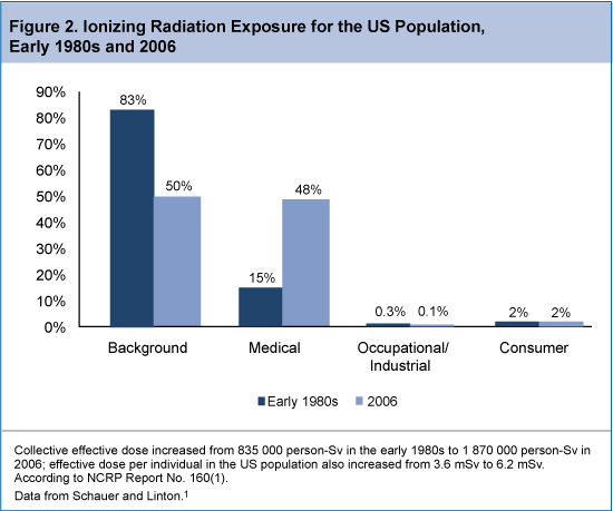
 The CT technologist should be aware of the radiation risks associated with CT, and can play a leadership role in implementing tools to limit radiation exposure in patients undergoing CT imaging. Specifically, CT technologists need to understand the parameters that affect radiation dose, practice dose reduction techniques on a regular basis, and be aware of the CT examination types that are the biggest contributors to radiation exposure from CT imaging. The increased use of CT in routine clinical practice has likely resulted in a substantial increase in radiation exposure in the US population. In fact, a report from the National Council on Radiation Protection and Measurements noted as much as 7-fold increase in ionizing radiation exposure from medical procedures between the early 1980s and 2006 (Figure 2). The report estimated that in 2006, 48% of total radiation exposure was from medical procedures, as opposed to just 15% in the early 1980s.13 Among the leading contributors to the increase in CT-associated radiation exposure include imaging studies of the head, chest, abdomen/pelvis, and studies of the chest for the diagnosis of pulmonary embolism.
The ongoing desire to limit radiation exposure must be balanced with the need to obtain adequate images during CT examinations. In general, higher radiation doses result in higher-resolution images, whereas lower doses lead to increased image noise and less sharp images. However, increased radiation dosage increases the risk of adverse side effects, most notably the risk of radiation-induced cancer. For instance, a 4-phase abdominal CT gives the same radiation dose as 400 chest X-rays. Fortunately, multiple strategies exist that can reduce the exposure to ionizing radiation during a CT scan.
Computed tomography technologists can employ a variety of dose reduction techniques that adequately balance the need for adequate imaging with adequate patient protection. For instance, technologists can control the z-axis scan length or minimize the number of phases to limit radiation exposure. External bismuth body shields to the breast, gonads, and thyroid are commonly used to protect these sensitive areas during CT imaging. In addition, dose reduction hardware techniques are commonly employed, including noise index and automatic tube current modulation, adjustments for peak kilovoltage (kVp), rotation time, and precise centering on the area to be scanned. In one analysis of the use of bismuth shielding, organ-based tube current modulation, and global tube current reduction as radiation dose reduction strategies using thorax phantoms of different sizes (15, 30, 35, and 40 cm lateral width), radiation dose to the breast region was reduced by approximately 21% and 37% with pediatric (2-ply shield; 15-cm phantom) and adult (4-ply shield; 30-, 35-, and 40-cm phantoms) models, respectively. Organ-based tube current modulation achieved dose decreases of 12% in the pediatric model and 34% to 39% in the adult model, and global lowering of the tube current reduced breast dosage by 23% and 39% in the pediatric and adult models, respectively.

The Importance of Patient Centering to Limit Radiation Dose in CT


The use of various tools that influence radiation dose and image noise, such as X-ray intensity shaping filters (bowtie filters), often requires accurate patient centering in the scan field of view. With the use of these bowtie filters, patient centering is critical to achieve the optimal balance of reduced radiation dose with reduced noise. To assess the influence of patient centering on image quality and dose requirements, researchers conducted imaging scans with bowtie filters using phantom centers that were positioned at 0, 3, and 6 cm below the center of rotation, or isocenter. These models were then retrospectively applied to scans from 273 adult body patients. The researchers reported that miscentering by 3 to 6 cm resulted in an increase in surface dose of 18% and 41%, respectively, and an increase in image noise of 6% and 22%, respectively. In addition, the retrospective analysis of adult body scout scan projection radiograph scans found that 46% of patients were miscentered in elevation by 20 to 60 mm, with a mean of 23 mm below the isocenter. Overall, the study concluded that patient miscentering resulted in a dose penalty of up to 140%, with a mean penalty of 33%, assuming that increases in tube current were necessary to compensate for increased image noise. The authors suggested automatically providing patient-specific centering and scan parameter selection information during CT studies so that radiologic technologists could improve workflow, achieve consistent image quality, and reduce radiation dose.19
Another study focused on the impact of patient miscentering on image noise and surface radiation dose using 3 different commercial CT scanners. Phantoms were positioned at 0, 2, 4, and 6 cm below the isocenter of the scanner's field of view. The resulting measurements were applied to imaging studies from 480 patients. The researchers reported that for a 64-slice CT scanner, the maximum increase in surface dose was 13.5%, 33.3%, and 51.1% with miscenterings of 2, 4, and 6 cm, respectively. An analysis of the available patient scout scans determined that patients were miscentered by an average of 2.2 cm below the isocenter. This resulted in increases in patient dose and image noise by 23% and 7%, respectively.20These studies demonstrate the importance of patient centering to limit radiation dose while optimizing image quality.

CT Parameters Affecting Radiation Dose


The discussion of strategies to estimate and limit radiation dose in patients undergoing CT will require a review of the specific CT parameters that most affect radiation dose, as defined in the sections below.8
Kilovolts
Kilovolt (kV) is the amount of voltage between an X-ray tube's anode and cathode. It determines the energy of the X-ray being emitted. Higher energy X-rays have a greater potential of passing through the body and creating a signal at the detector than lower energy X-rays. Higher kV also means less noise. High-energy X-rays are absorbed by the body and deposit more energy than low-energy X-rays; therefore, contribute more to patient dose. For example, changing the kV from 120 to 135 increases the radiation dose delivered by approximately 33%. However, higher kV is sometimes warranted in larger patients or dense anatomy such as bone where increased penetration is needed.
MilliAmpere SecondsThe tube current or milliAmpere seconds (mAs) determines the number of X-rays the tube produces. Combined with the gantry rotation time, mAs represents the total X-ray output of the tube per rotation. Changing the mAs, is the most common method of adjusting dose and noise level. Cutting the mAs in half will reduce the dose by a factor of 2; however, it will also half the number of X-rays reaching the detector. Because fewer X-rays are detected, the image noise will increase and low contrast detectability will be diminished.
CT Pitch and Helical Pitch-Beam Pitch
CT pitch and helical pitch-beam pitch is defined as the distance the table travels in a rotation divided by the total active detector width in the Z direction. Helical pitch is the same except it is divided by the individual channel thickness rather than the total collimation. The higher the CT helical pitch, the faster the table moves through the X-ray beam. This results in lower radiation dose to the patient, however it may result in increased noise and may cause helical artifact. One may need to increase mAs as the pitch increases to maintain image quality.
Effective mAs Effective mAs (mAs eff) is the mAs divided by the pitch. Because pitch affects the patient dose, mAs by itself does not completely represent the number of X-rays entering the patient.
CollimationWith multislice scanning, there are many combinations of slice width and the number of slices that may be used to acquire the scan volume. With all collimators on multislice systems, the actual X-ray beam is slightly wider than the nominal beam width. This is to ensure that the detectors on the edge of the array receive uniform X-ray coverage. This results in a small amount of unused radiation. For the best image quality and dose efficiency, using the thinnest slices to cover the entire detector is ideal.
Acquired and Reconstructed Slice WidthAcquired is the actual slice thickness at which the scan was taken. Reconstructed slice width refers to post processing of the acquired slice. The acquired slice width is governed by the multislice detector configuration and determines the minimum image width that can be reconstructed. Thicker reconstructed images have less noise with all other factors being equal. However, thicker slices have less resolution. Scanning with the thinnest possible slice avoids partial volume artifacts and allows flexibility for excellent multiplanar reformats and 3-dimensional rendering without higher radiation dose. The optimal image quality and dose efficiency are achieved with the thinnest slice covering the entire detector.
Reconstruction KernelThe acquired data are filtered by the reconstruction kernel. The kernel plays a large role in determining spatial resolution. This has a great effect on the amount of noise in the image and the dose needed for a given level of image noise. There are a large variety of reconstruction kernels to select. The choice of the kernel is made based on the clinical need. Sharper reconstruction kernels are used for better in-plane spatial resolution such as in the lungs to help identify tiny airways. Smooth reconstruction kernels reduce the image noise at the expense of in-plane resolution; they are used in body imaging to reduce noise and enhance contrast resolution.
Detector Efficiency
Detector efficiency has a huge influence on the radiation dose delivered during a CT examination. The detector's ability to capture the X-ray and convert it to light, then transmit that light and convert it into an electrical signal with minimal loss, defines the overall efficiency of the detector. Better efficient detectors result in lower patient doses for a given level of image quality.
FiltrationFiltration is added outside the X-ray tube to block low-energy X-rays (photons) and reduce patient dose. The lowest energy photons will not pass through the body at all and will only contribute to patient dose. In the process of removing these low-energy photons, some desirable medium- and high-energy X-rays will be removed as well. This results in decreasing overall output of the X-ray tube. An increase in tube current or mAs may be needed to maintain image quality. A CT system needs enough filtration to block the lowest energy photons but not so much to lose the ability to distinguish low-contrast anatomy.
Patient SizeThe size of the patient plays a significant role in the total dose absorbed for the same technique. It is important to tailor the kV and mAs to the patient size to minimize the dose to the patient. With smaller patients, such as pediatric patients, the dose can be 2 to 3 times higher than on an adult. Lower kV and mAs should be used to achieve the small image quality.
Body Part Being ScannedDifferent organs in the body have different sensitivities to radiation (Table 1). For instance, the lungs are not as dense as the abdomen or pelvis because they are air filled. Therefore, less radiation (kVp and mA) is needed for imaging.
AgeThe risk of developing cancer from a CT examination decreases with increased age. Younger patients' organs are more radiosensitive in general, due to the rapid rate of cell division and growth at a young age. It is critical in young patients to keep the dose as low as possible while maintaining image quality.

How radiation affects cells


The primary way radiation affects our health is through breakage of DNA molecules. DNA is a long chain of amino acids whose pattern forms the blueprint on how the cell lives and functions.  Radiation is able to break that chain. When it does,  three things can happen:  

1) The DNA is repaired properly

In this case, the cell is repaired properly and it continues to function normally. DNA breakage occurs normally every second of the day and cells have a natural ability to repair that damage.

2) The DNA damage is so severe that the cell dies (deterministic effects)

When the DNA or other critical parts of a cell receive a large dose of radiation, the cell may either die or be damaged beyond repair. If this happens to a large number of cells in a tissue or organ, early radiation effects may occur. These are called deterministic effects and the severity of the effects varies according to the radiation dose received. They can include burns, cataracts, and in extreme cases, death.

The first evidence of deterministic effects became apparent with early experimenters and users of radiation. They suffered severe skin and hand damage due to excessive radiation dose. More recently, this relationship was observed at the 1986 Chernobyl nuclear plant accident where more than 130 workers and firefighters received high radiation doses (800 to 16,000 mSv), and suffered severe radiation sickness. Two of the people exposed died within days of exposure. Close to 30 more workers and firefighters died within the first three months.

The CNSC and other international regulators put measures in place, including stringent dose limits and radioactive source tracking databases, to mitigate the chances of the public or workers receiving doses of radiation high enough to cause deterministic effects. The CNSC also has strict regulations on how nuclear substances and devices must be handled in Canada. 
3) The cell incorrectly repairs itself, but it continues to live (stochastic effects)

In some cases, the DNA of the cell may be damaged by radiation, but the damage does not kill the cell.  The cell may continue to live and even reproduce itself, but the cell and its descendents may no longer function properly and may disrupt the function of other cells.  The probability of this type of  detrimental effect is proportionate to the dose and it is called a stochastic effect – when there is a statistical probability that the effects of exposure will occur. In such cases, the likelihood of the effects increases as the dose increases. However, the timing of the effects or their severity does not depend on the dose. 

This process happens all the time in everyone. In fact, people are exposed to about 15,000 such events every second of every day. Sometimes, the cell structure changes because it repairs itself improperly. This alteration could have no further effect, or the effect could show up later in life. Cancer and hereditary effects may or may not take place.

Radiation-induced hereditary effects


Genetic damage occurs when the DNA of sperm or egg cells are damaged. This causes a harmful characteristic that is passed on from one generation to the next. Animal studies, such as those conducted on fruit flies by Hermann J. Muller in 1926, showed that radiation will cause genetic mutations. However, to date there have been no known genetic effects in humans caused by radiation. This includes studies involving some 30,000 children of survivors of the atomic bombings of the cities of Hiroshima and Nagasaki in Japan in 1945 (BEIR VII).

Popular Posts