In early 2011, the US Food and Drug Administration (FDA) approved the first cardiac pacemaker designed to be used safely during magnetic resonance imaging (MRI) examinations.
The Revo MRI SureScan pacing system is MR Conditional designed to allow patients to undergo MRI under the specified conditions for use. A complete system, consisting of a Medtronic Revo MRI SureScan IPG implanted with two CapSureFix MRI™ SureScan leads is required for use in the MRI environment.
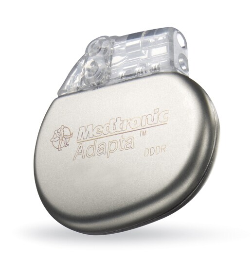
. Revo MRI™ SureScan® Pacing System
Design Features of MRI-safe pacemaker Revo MRI SureScan
The Revo MRI SureScan Pacing System is based on the Medtronic EnRhythm pacemaker and CapSureFix Novus pacing lead, with the following modifications to improve MRI compatibility :
The Revo MRI SureScan pacing system is MR Conditional designed to allow patients to undergo MRI under the specified conditions for use. A complete system, consisting of a Medtronic Revo MRI SureScan IPG implanted with two CapSureFix MRI™ SureScan leads is required for use in the MRI environment.
.
Revo MRI™ SureScan® Pacing System Design Features of MRI-safe pacemaker Revo MRI SureScan
The Revo MRI SureScan Pacing System is based on the Medtronic EnRhythm pacemaker and CapSureFix Novus pacing lead, with the following modifications to improve MRI compatibility :
- Modified leads to reduce RF-induced lead tip heating
- Internal circuits changed to reduce the potential for cardiac stimulation
- Amount of ferromagnetic materials limited
- Internal circuit protection improved to prevent disruption of internal power supply
- Pacemaker reed switch replaced with a Hall sensor, with predictable behavior in a static magnetic field
Specifications: of MRI-safe pacemaker-Revo MRI SureScan
| Revo MRI SureScan Pacemaker | |
|---|---|
| Size | 12.7 cc |
| Weight | 21.5 g |
| CapSureFix MRI Model 5086 | |
| Polarity | Bipolar |
| Shape | Straight |
| Fixation | Screw-in |
| Inner/Outer Insulator | Silicone |
| Body | 7 Fr |
| Recommended Introducer Size | 8 Fr (without guide wire) |
| Tip-to-Ring Spacing | 11 Fr (with guide wire) |
| Standard Lengths | 45 cm, 52 cm, 58 cm |
Indications MRI-safe pacemaker-Revo MRI SureScan
It is estimated that up to 75% of pacemaker patients will have a medical need for an MRI over the lifetime of their device.
MRI has increasingly become the diagnostic imaging modality of choice for many disease states, including cancer, stroke, dementia, and musculoskeletal disorders. Such disorders are not uncommon among patients with a pacemaker or an ICD who require treatment; it has been estimated that a patient with an implanted cardiovascular device has at least a 50% likelihood of having a clinical indication for MRI over the lifetime of the device.
Electromagnetic fields and radiofrequency (RF) energy generated by an MRI scanner may pose risks to patients with a pacemaker, such as interference with pacemaker operation, damage to system components, inappropriate therapy, lead or pacemaker dislodgement, or change in pacing capture threshold. A scientific statement from the American Heart Association (AHA) on the safety of MRI in patients with cardiovascular devices includes limited recommendations on the performance of MRI in patients with a pacemaker/ICD. These include the following:
- Performing the exam at centers with expertise in MRI and electrophysiology
- Having a physician with pacemaker/ICD expertise decide whether it is necessary to reprogram the device before the MRI exam
- Having a person with expertise in MR physics and safety involved in planning the scan, with consideration for the use of scanning parameters that minimize risk (eg, lowest RF power levels, weakest/slowest necessary gradient magnetic fields)
- Testing pacemaker functions before and after the exam
- Observing the patient closely throughout the exam, including monitoring of heart rhythm and vital signs
Nevertheless, considerable controversy has remained over safety issues. MRI is generally contraindicated in patients with pacemakers, and the AHA guidelines recommend consideration of MRI only in exceptional circumstances, excluding the vast majority of pacemaker patients who might benefit from MRI examination.
In February 2011, the FDA approved the Revo MRI SureScan Pacing System, the first cardiac pacemaker designed to be used safely during MRI exams.
Clinical Trial Evidence of MRI-safe pacemaker-Revo MRI SureScan
No MRI-related complications occurred during or after MRI in an investigation of the safety and effectiveness of the Revo system, the EnRhythm MRI SureScan Pacing System Study. This prospective, randomized, controlled, multicenter clinical trial examined patients who met class I or II dual-chamber pacemaker implant indications according to the American College of Cardiology/American Heart Association/Heart Rhythm Society (ACC/AHA/HRS) 2008 Guidelines for Device-Based Therapy of Cardiac Rhythm Abnormalities.
Patients were randomized to undergo an MRI scan 9-12 weeks after pacemaker implantation (MRI group, n = 258) or not to undergo MRI (control group, n = 206). MRI scans were performed on 1.5T systems from 3 different manufacturers. Fourteen nonclinically indicated head and lumbar spine sequences were performed on each patient in the MRI group. As stated above, no MRI-related complications occurred during or after MRI, including sustained ventricular arrhythmias, pacemaker inhibition or output failures, electrical resets, or threshold or sensitivity changes.
Clinical Implementation of MRI-safe pacemaker-Revo MRI SureScan
A dedicated programming care pathway was developed for the (MRI)-safe pacemaker (ie, the Revo MRI SureScan Pacing System) to facilitate the choice between asynchronous and nonstimulation modes, increase the pacing output to 5.0 V/1.0 ms during MRI, prevent programming of the MRI mode if the device has failed any of the 7 system integrity checks (see below), and facilitate restoration of prescan program states and values.
A dedicated programming care pathway was developed for the (MRI)-safe pacemaker (ie, the Revo MRI SureScan Pacing System) to facilitate the choice between asynchronous and nonstimulation modes, increase the pacing output to 5.0 V/1.0 ms during MRI, prevent programming of the MRI mode if the device has failed any of the 7 system integrity checks (see below), and facilitate restoration of prescan program states and values.
Pacing system integrity checks are as follows:
- Pacemaker and both leads implanted for more than 6 weeks
- Pectoral implantation
- No other active pacing, ICDs, or leads
- No abandoned leads, lead extenders, or adapters
- Leads electrically intact, with stable and normal function
- Lead impedance between 200 and 1500 W
- Capture threshold less than 2.0 V at 0.4 ms
Follow-up/Monitoring
Recommended follow-up visits should be scheduled (as for other device or lead implantations) as follows:
Recommended follow-up visits should be scheduled (as for other device or lead implantations) as follows:
- 1-2 weeks - Wound check
- 1 month - Pacemaker interrogation
- 3 months - Pacemaker interrogation
- Every 6 months thereafter - Pacemaker interrogation
As with other pacemakers, an identification card is provided to patients implanted with the Revo pacemaker. This information is crucial when communicating with the cardiologist about a pacer problem. Like other pacemaker systems, the Revo system includes radiopaque markers that can be seen on a standard chest radiograph. These identification markers are placed on pacemakers and leads.
Medtronic first-generation Revo MRI SureScan Pacing System has some significant limitations.
1. The MRI-pacemaker is for new heart patients. Patients who already have a pacemaker can not get this new model unless they undergo the risky procedure of having their old pacemaker completely removed. Usually, when it comes time to replace the battery in a pacemaker (about 5-7 years), the metal case containing battery and circuitry is detached from the leads, and a new model device is hooked up to the leads. But doctors generally consider it too risky to remove the old leads from the heart for fear of tearing the heart or the veins through which the leads are inserted into the heart. Part of the design of the Revo pacemaker is its new leads and so they must be the leads that connect the pacemaker to the patient’s heart.
2. Patients must have the Revo pacemaker implanted for 6 weeks before receiving an MRI.
3. The Revo pacemaker requires a certain position of the patient inside the MRI tube so as to avoid most chest scans. This is to prevent overheating the metal tips of the leads that are attached to the heart. So heart scans are forbidden with this first generation model.
4. And Owen Faris, senior scientific reviewer for the FDA, explains that the new pacemaker won’t work for all types of MRI scans and won’t work in all MRI scanners. In his words:
In addition to the chest scan exclusion, there is a restriction on how much radio-frequency energy can be deposited into the body by the scanner. MRI scanners have two operating modes for most clinical applications. ‘Normal operating mode’ is how the scanner is normally programmed and that mode restricts the scanner to lower-energy scans (less than 2 Watts per kilogram). This is sufficient energy for most clinical MRI scans. However, for some patients and for certain scans, more power is needed. In those cases, the MRI scanner is placed in ‘First level control’ mode, which allows for greater energy deposition (up to 4 Watts per kilogram). For patients implanted with the REVO MRI pacemaker, those patients are not allowed to have these higher energy scans.
MRIs for these patients are also restricted to only allow use of 1.5 tesla MRI systems. "Tesla" is a measure of the strength of the magnetic field.
5. Medicare does not now pay for MRI scans on a patient who has a pacemaker. Medtronic spokesperson Wendy Dougherty says that the company will not speculate on whether Medicare will cover MRIs done on patients wearing the new Revo pacemaker. The federal agency is considering a petition from a physician to cover MRIs done during an investigational study to determine the risk of MRIs involving pacemakers already in use. During the comment period for this request, Medtronic asked Medicare to restrict MRI payment to patients wearing pacemakers approved by the FDA for use with MRIs. Medicare’s decision is due by March 1. Patients on Medicare would be wise to check on whether their plan will pay for an MRI before getting the test, which costs between $1,600 and $3,500 at different medical centers and offices.


